
Recent work in the Fiore lab (https://biology.appstate.edu/directory/dr-cara-fiore) has used scanning electron microscopy (SEM) for identification of freshwater sponges using skeletal structures called spicules. The spicules are silica-based structures with diverse morphology (Figure 1). These structures vary across sponge species and are critical for taxonomic identification of the sponges. We also plan to use transmission electron microscopy (TEM) to determine the density, type, and localization of microbial symbionts in marine and freshwater sponges. The freshwater sponge system is unique and one that our lab is developing into an experimental host-microbe system. Specifically, we see that the sponge microbiome changes over the life span of the sponge from a community identical to that of the surrounding water column community to one that is distinct from that of the water, suggesting some host selection of symbiotic bacteria. We will use several types of microscopy to identify the types of symbiotic bacterial present and identify potential structures for interaction between bacterial symbionts and the sponge host.
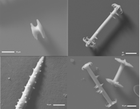
Figure 1. Scanning electron microscopy images of freshwater sponge spicules.